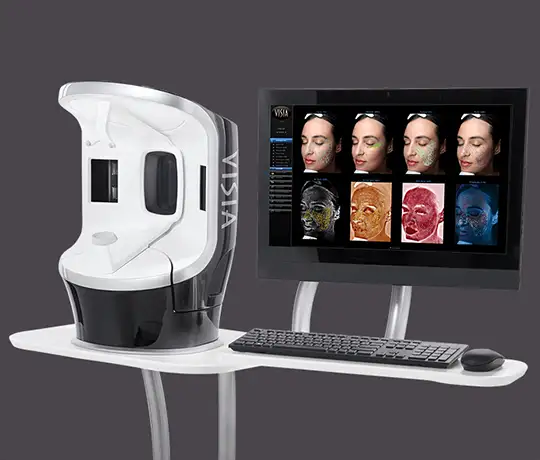
Visia 3D Professional Skin Analysis
Visia 3D AI Digital Skin & Complexion Analysis
Clinical Diagnostic Summary: The Visia 3D Skin Analysis system is an AI-powered, multi-spectral digital diagnostic platform that quantit...
Clinical Diagnostic Summary: The Visia 3D Skin Analysis system is an AI-powered, multi-spectral digital diagnostic platform that quantitatively maps both epidermal surface conditions and subsurface dermal architecture. Utilized under the medical supervision of Prof. Dr. Ismail Kucuker at our Istanbul clinic, Visia harnesses cross-polarized, non-polarized, and UV lighting to objectively score UV solar damage, melasma, wrinkle depth, pore caliber, vascular inflammation, and bacterial porphyrins against the world's largest dermatological database—establishing your biological TruSkin Age® without guesswork.
Cumulative ultraviolet radiation, atmospheric toxins, metabolic stress, and biological chronaging compromise the skin's cellular matrix long before structural damage manifests visible symptoms in a conventional mirror.
At Prof. Dr. Ismail Kucuker Clinic in Istanbul, clinical excellence begins with precision diagnostics. The Visia 3D multi-spectral optical system eliminates trial-and-error by capturing high-definition cross-sectional imagery of both surface and deeper subcutaneous planes.
Plastic, Reconstructive and Aesthetic Surgeon Prof. Dr. Ismail Kucuker analyzes these algorithmic indicators to design targeted, multi-modality treatment regimens customized strictly to your biological tissue requirements.

Visia's proprietary multi-spectral algorithms isolate, evaluate, and rank 8 critical dermal variables against age- and skin-matched clinical cohorts:
The diagnostic session takes place within an optically controlled capture booth. An automated rotational sensor glides seamlessly to record high-resolution images from three standardized angles: left profile, frontal, and right profile.
By synchronizing IntelliFlash® cross-polarized filters with diagnostic UV illumination, the system separates color signatures to reveal underlying cellular behavior. Canfield's software immediately contrasts your personal findings against a global peer database of identical biological age, gender, and Fitzpatrick phototype to establish your definitive TruSkin Age®.
Precision Protocol Selection: Rather than applying generic treatments, diagnostic data directly governs procedural choice at our clinic. Severe subsurface vascular congestion directs protocols toward restorative cellular therapies, while isolated melanin sheets are targeted with precise fractional laser wavelengths or medical chemical peels.
Longitudinal Digital Validation: During follow-up visits, newly captured scans are digitally superimposed over your pre-treatment baseline. This mathematically confirms reduction in pigment density, thickening of collagenous networks, and tightening of pore calibers, ensuring transparent, accountable aesthetic care.
Medical Notice: The medical information, surgical recovery outlines, and treatment descriptions presented on this website are intended strictly for educational and informational purposes. They do not constitute formal medical diagnosis, individualized healthcare advice, or surgical guarantees. Final candidacy and custom surgical planning are determined exclusively during an in-person clinical consultation with a board-certified plastic surgeon.